

The global market for diagnostic imaging is projected to reach $7.4 billion by September 2026, yet the technical complexity of selecting the right mri machine remains a significant hurdle for most healthcare administrators. You understand that field strength selection and facility shielding requirements represent high-stakes decisions that directly impact both clinical outcomes and institutional financial stability.
This authoritative guide provides a comprehensive framework for understanding modern MRI technology, from Tesla ratings to the logistical demands of facility preparation. We'll analyze the specific clinical applications of 1.5T and 3.0T systems while detailing the procurement requirements for healthcare facilities in September 2026. By examining current regulatory standards and the latest advancements in AI-enhanced imaging, you'll gain the clarity needed to mitigate capital expenditure risks. This overview ensures your facility maintains operational continuity while meeting the rigorous demands of modern diagnostic medicine through precise, data-driven equipment selection.
Key Takeaways
- Learn the fundamental science behind Magnetic Resonance Imaging (MRI), including how hydrogen atom alignment and radiofrequency pulses generate high-resolution diagnostic data.
- Identify the critical differences between Tesla (T) ratings to select the most appropriate mri machine for your facility's clinical workload and patient throughput.
- Explore diverse clinical applications, ranging from advanced neurological stroke detection to detailed musculoskeletal evaluations of ligament and cartilage health.
- Master essential facility requirements for September 2026, including the installation of a Faraday Cage and specialized magnetic shielding to ensure institutional safety compliance.
- Navigate the 2026 procurement landscape by evaluating the Total Cost of Ownership (TCO), including maintenance, power consumption, and the strategic benefits of accredited refurbished equipment.
The Mechanism of Magnetic Resonance Imaging (MRI)
Magnetic Resonance Imaging (MRI) is a non-invasive diagnostic technique that utilizes powerful magnetic fields and radio waves to generate detailed anatomical images. Unlike Computed Tomography (CT) or X-ray systems, an mri machine does not rely on ionizing radiation. This provides a significant safety profile for patients requiring repeated scans. The Mechanism of Magnetic Resonance Imaging relies on the interaction between the equipment's magnetic field and the body's internal chemistry to provide superior soft-tissue contrast. This capability is vital for identifying pathologies in the brain, spinal cord, and muscles that other imaging modalities might miss.
To better understand this concept, watch this helpful video:
Nuclear Magnetic Resonance and Hydrogen Atoms
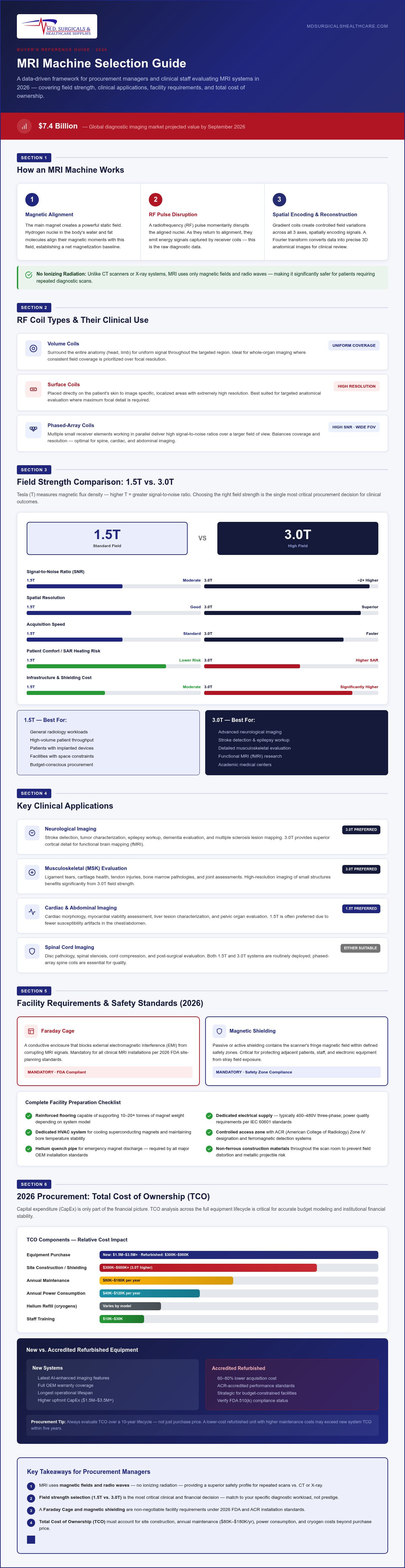
Hydrogen is the primary target for medical imaging because it's the most abundant element in the human body, specifically within water and fat molecules. The main magnet of the mri machine creates a static, uniform magnetic field that aligns the magnetic moments of these hydrogen nuclei. In the context of MRI physics for September 2026, proton spin refers to the intrinsic angular momentum of hydrogen nuclei that allows them to interact with external magnetic fields. When you place a patient in the bore, these spins align with the field, creating a net magnetization. This alignment serves as the baseline for all subsequent diagnostic data collection.
Radiofrequency Pulses and Gradient Coils
Once the atoms are aligned, the system applies a radiofrequency (RF) pulse to disrupt this state momentarily. As the nuclei return to their original alignment, they emit energy that the system captures as electrical signals. Gradient coils are essential for localizing these signals within the body by creating controlled, linear variations in the magnetic field strength across the three axes. This spatial encoding allows the reconstruction of data into precise three-dimensional images. The captured energy is processed via a Fourier transform to create the final visual output used by clinicians.
RF coils function as the antennas of the system, transmitting the pulse and receiving the resulting signal. You'll select different coils based on the specific clinical application:
- Volume coils: These surround the entire anatomy, such as the head or a limb, to provide a uniform signal throughout the targeted area.
- Surface coils: These are placed directly on the patient's skin to image specific, localized areas with extremely high resolution.
- Phased-array coils: These utilize multiple small receiver elements to provide high signal-to-noise ratios over a larger field of view.
The choice of coil directly impacts image quality and diagnostic accuracy for your clinical staff. Selecting the appropriate hardware ensures that the emitted energy is captured with maximum efficiency, reducing the need for repeat scans and improving patient throughput.
Technical Specifications for 2026 MRI Systems
Selecting an mri machine requires a granular understanding of field strength, which is measured in Tesla (T) units. This measurement represents the density of the magnetic flux and determines the signal-to-noise ratio available for imaging. Understanding How MRI scanners work helps procurement teams evaluate how these field strengths interact with biological tissues to produce diagnostic data. Beyond the magnet itself, you must evaluate the slew rate and gradient strength, as these specifications directly dictate acquisition speed and image resolution.
Bore size is another critical specification for patient comfort and clinical throughput. Standard 60 cm bores are increasingly being replaced by 70 cm wide-bore systems to accommodate bariatric patients and reduce claustrophobia. Some specialized models, such as the Siemens MAGNETOM Free.Max, now feature an 80 cm bore. These wider apertures significantly decrease the rate of aborted scans, ensuring more reliable scheduling and improved patient experiences across your facility.
Comparing 1.5T and 3.0T Field Strengths
The 1.5T MRI remains the global clinical standard in September 2026, accounting for over 80% of routine diagnostic scans. It offers exceptional stability for orthopedic and general body imaging while remaining less sensitive to susceptibility artifacts caused by metal implants. In contrast, 3.0T MRI systems provide double the signal-to-noise ratio, making them superior for advanced neuroimaging and vascular studies. In the 2026 market, a new unit typically ranges from $1,000,000–$3,000,000 depending on the specific configuration and software packages included.
Emerging 7.0T Systems and Ultra-High Field Imaging
Ultra-high field systems, specifically 7.0T MRI, have transitioned from purely research environments into specialized clinical use for complex neurological applications. These machines offer unparalleled Signal-to-Noise Ratio (SNR), allowing for metabolic imaging and the visualization of microstructures in the brain that are invisible at lower field strengths. However, they present significant logistical challenges, including extensive magnetic shielding and dedicated facility structural reinforcements. As of September 2026, these remain high-capital investments primarily reserved for tertiary care centers and academic research institutions.
The September 2026 market is also defined by the shift toward Helium-free cooling and AI-enhanced reconstruction. Conventional scanners require over 1,000 liters of liquid helium, but newer platforms like the Siemens MAGNETOM Flow require as little as 0.7 liters. This technology mitigates the risk of helium supply chain volatility and reduces long-term maintenance costs. Additionally, AI-driven software, such as GE's AIR Recon DL, now reduces scan times by up to 50% without compromising image quality. If you're evaluating these specifications for your facility, you can consult with our procurement experts to verify current model availability and logistical requirements.
Clinical Applications and Diagnostic Efficacy
The clinical versatility of a modern mri machine extends far beyond simple anatomical visualization. In September 2026, healthcare providers utilize these systems for complex neurological diagnostics, including the early detection of ischemic strokes and the monitoring of multiple sclerosis (MS) progression. High-resolution imaging allows clinicians to distinguish between benign and malignant tumors with exceptional precision. Beyond the brain, Musculoskeletal (MSK) imaging remains a high-volume application. It's the primary modality for evaluating ligament tears, cartilage damage, and chronic spinal health issues. The diagnostic accuracy provided by these scans reduces the need for invasive exploratory procedures.
Abdominal and Pelvic Imaging
MRI provides a distinct advantage over Computed Tomography (CT) for pelvic soft tissue visualization due to its superior contrast resolution. In oncology, it's indispensable for the precise staging of liver, pancreas, and prostate cancers. Clinicians often use contrast agents like Gadolinium to enhance diagnostic clarity by highlighting vascularity and tissue permeability. This level of detail is critical for surgical planning and monitoring treatment response in complex oncology cases. The ability to visualize narrow anatomical structures without ionizing radiation makes it the preferred choice for pediatric and long-term care patients.
Cardiac MRI and Vascular Studies
Cardiovascular assessment has seen significant growth as a non-invasive alternative to catheterization for certain pathologies. Cardiac MRI allows for the detailed evaluation of heart wall thickness, chamber function, and overall myocardial viability. It's particularly effective for managing congenital heart disease and identifying areas of scarring after a myocardial infarction. For vessel mapping, Magnetic Resonance Angiography (MRA) provides a non-invasive method to visualize arterial and venous structures. These sequences are often integrated with advanced patient monitors during stress-test sequences to ensure hemodynamic stability.
Functional MRI (fMRI) has also expanded from research into clinical practice for mapping brain activity. It measures hemodynamic changes in blood flow to identify active regions during specific tasks. This technique is vital for pre-surgical brain mapping to protect eloquent areas during tumor resection. By utilizing these advanced sequences, your clinical staff can achieve more confident diagnoses across a wider range of patient types. The multifaceted nature of the mri machine ensures it remains the cornerstone of a comprehensive diagnostic department in September 2026.
Facility Requirements and Safety Standards
Integrating a high-field mri machine into a clinical environment requires meticulous site preparation that goes beyond standard construction. You must install a Faraday Cage, which is an essential copper or aluminum radiofrequency (RF) shield that prevents external signals from contaminating diagnostic data. Simultaneously, magnetic shielding is required to contain the fringe field, protecting sensitive medical equipment and personnel in adjacent rooms. According to the March 2026 American College of Radiology (ACR) updates, facilities must strictly maintain a four-zone access model. Zone one is for general public access, while Zone two serves as the unscreened patient waiting area. Zone three is a restricted area for screened patients and personnel, and Zone four is the actual scanner room where the magnet is located.
Cryogen management remains a critical safety priority for superconducting magnets that utilize liquid helium for cooling. You must install a quench pipe, a specialized exhaust system designed to vent helium gas safely outside the building if the magnet loses its superconductivity. While newer systems in September 2026 utilize low-helium technology, traditional scanners still require robust protocols to prevent oxygen displacement within the scan room. Ensuring the structural integrity of this exhaust system is a non-negotiable requirement for regulatory compliance and institutional safety.
Site Preparation and Structural Engineering
Structural integrity is a primary concern because a typical magnet assembly can weigh between 10 and 25 tons. This necessitates reinforced flooring and specific weight-bearing assessments from structural engineers before installation begins. Urban clinics located on higher floors must also account for vibration isolation to prevent external transit or construction noise from degrading image quality. You'll also need dedicated electrical substations and high-capacity cooling systems to handle the significant power draw and heat dissipation required by the gradient coils during continuous operation.
Safety Protocols for Staff and Patients
Clinical safety protocols are built around rigorous screening processes for all individuals entering the facility. Staff must verify that patients don't have ferromagnetic implants or medical devices that could be dislodged or heated by the magnetic field. During high-gradient sequences, acoustic noise can exceed 100 decibels, making high-quality ear protection a standard requirement for patient safety and comfort. Emergency procedures must be clearly documented and practiced, including specific training for magnet quenching and managing power failures to maintain cryogen stability and patient security.
Strategic Procurement: Sourcing MRI Equipment
Procuring an mri machine involves more than the initial capital outlay. You must evaluate the Total Cost of Ownership (TCO), which includes annual service contracts ranging from $50,000–$150,000. These costs cover preventative maintenance, software patches, and emergency repairs necessary for operational continuity. Additionally, power consumption and specialized staffing requirements for Level 1 and Level 2 ACR-trained personnel represent significant ongoing operational expenses that impact your long-term budget.
The September 2026 market offers a robust selection of both new and accredited refurbished equipment. While a new 1.5T system may cost up to $2,000,000, a refurbished unit typically ranges from $350,000–$850,000. These pre-owned systems often provide savings of 40% to 60%, allowing facilities to expand their diagnostic capabilities without excessive financial strain. However, you'll need to ensure that any used hardware undergoes rigorous verification to meet current FDA and international safety standards.
Maximizing ROI in Diagnostic Imaging
Analyzing patient throughput is essential for calculating accurate reimbursement rates in September 2026. Selecting modular systems allows your facility to implement future software upgrades, such as AI-driven reconstruction, without replacing the entire magnet assembly. This approach extends the lifespan of your investment and ensures your clinical staff has access to the latest diagnostic tools. Rigorous procurement verification ensures that every piece of imaging hardware meets the precise regulatory standards required for hospital compliance and patient safety.
Partnering with M.D. Surgical & Healthcare Supplies
We provide access to a global network of verified diagnostic imaging hardware, ensuring that your procurement process is secure and efficient. Our team coordinates the complex logistics for MRI systems, CT scan machines, and necessary auxiliary equipment like patient monitors. We manage everything from international shipping to the final logistical verification at your facility site. You can contact our specialists for customized institutional quotes and detailed shipping timelines tailored to your specific project requirements.
Advancing Diagnostic Capabilities in September 2026
Selecting a high-capacity mri machine requires a balanced assessment of technical specifications, clinical diagnostic goals, and long-term facility requirements. You've explored how field strengths like 1.5T and 3.0T dictate image resolution and how rigorous adherence to ACR safety zones protects both your staff and patients. Strategic procurement in September 2026 demands meticulous verification of equipment history and a clear understanding of the total cost of ownership to mitigate financial risk.
M.D. Surgical & Healthcare Supplies operates as a licensed global distributor focused on the secure delivery of high-value imaging technology. Our dedicated procurement specialists provide institutional buyers with full logistical verification and transparent shipping protocols for every unit. We anticipate supply chain gaps to ensure your facility maintains operational continuity without compromising on clinical precision. Our role is to serve as your dependable logistical backbone in an increasingly complex healthcare environment.
We look forward to partnering with your institution to enhance your diagnostic infrastructure and patient care standards with unwavering stability and professional excellence.
Frequently Asked Questions
What is the difference between a closed and wide bore MRI machine?
A standard closed bore system typically features a 60 cm diameter aperture. In contrast, a wide bore mri machine offers a 70 cm or 80 cm opening. This increased space significantly improves patient comfort and reduces the incidence of claustrophobia. It also allows clinical staff to accommodate bariatric patients more effectively. Facilities often prioritize wide bore designs to decrease the rate of aborted scans and improve overall institutional throughput.
How much does a new MRI machine cost in 2026?
In September 2026, the price for a high-end mri machine from a licensed distributor typically falls within the range of $50,000–$500,000+. This pricing depends on the specific configuration, field strength, and software packages included. Sourcing through a global network ensures that all imaging hardware is accredited and verified before sale. Procurement managers should also account for additional costs related to site preparation, such as RF shielding and structural reinforcements for the magnet magnet.
Is an MRI scan safer than a CT scan regarding radiation?
An MRI scan is generally considered safer regarding radiation because it utilizes non-ionizing magnetic fields and radio waves. Unlike a CT scan, which relies on ionizing X-ray radiation, magnetic resonance imaging does not damage DNA or increase long-term cancer risks. This makes it the preferred diagnostic tool for pediatric patients and individuals requiring frequent follow-up imaging. Clinical staff must still follow strict safety protocols to manage the powerful static magnetic field within the facility.
What are the primary power requirements for a 3.0T MRI system?
A 3.0T MRI system requires a highly stable, industrial-grade electrical supply to function. Most systems necessitate a dedicated substation and a 480V three-phase power connection to handle peak loads during gradient-heavy sequences. You'll also need a high-capacity water-cooling system, or chiller, to dissipate the heat generated by the gradient coils. Proper electrical conditioning is essential to prevent power fluctuations from degrading image quality or damaging sensitive electronic components within the scanner.
How long does a standard MRI imaging sequence take?
A standard imaging sequence typically lasts between 15 and 90 minutes. The exact duration depends on the body part being scanned and the number of sequences required for a definitive diagnosis. In September 2026, AI-powered reconstruction technologies like Deep Learning can reduce these times by as much as 50%. This advancement allows for higher patient turnover while maintaining the superior soft-tissue contrast that clinical staff require for accurate neurological and musculoskeletal assessments.
What is the role of Gadolinium in an MRI contrast study?
Gadolinium is a rare-earth metal used as a contrast agent to improve diagnostic clarity. It works by temporarily altering the magnetic properties of water molecules in the body, which enhances the signal in specific tissues. This allows radiologists to identify tumors, inflammation, and blood supply issues with greater precision. While generally safe, clinical staff must screen patients for renal function before administration to ensure the contrast agent is cleared safely from the system.
Can patients with metal implants undergo an MRI scan?
Many patients with metal implants can safely undergo a scan if their device is labeled as MRI Conditional. This means the implant has been tested and found safe under specific magnetic field parameters. However, patients with older pacemakers, cochlear implants, or certain ferromagnetic clips may be restricted from entering Zone four. Clinical staff must perform a rigorous screening process to identify any internal hardware and verify its compatibility with the specific field strength of the scanner.
How often does an MRI machine require cryogen refilling?
The frequency of cryogen refilling depends on the magnet's design and cooling technology. Traditional superconducting magnets require liquid helium refills periodically to maintain their magnetic field. However, many systems in September 2026 utilize zero-boil-off technology or ultra-low helium designs that rarely require refilling. These advancements mitigate the risks associated with helium supply chain volatility. They also reduce the long-term maintenance costs for healthcare facilities by minimizing the need for manual cryogen management.

