

Selecting the highest field strength available isn't always the smartest financial or clinical move for a modern healthcare facility. Mastering the nuances of mri machine specifications is the difference between acquiring a high-utility diagnostic tool and a costly logistical burden. You're likely facing a wall of technical jargon like Slew Rate and Tesla while worrying about over-investing in features your patient volume doesn't justify. It's a common concern that can lead to significant budgetary waste if not addressed with technical precision and market awareness.
This guide simplifies these complex parameters to help you secure a system that maximizes both clinical outcomes and return on investment. We'll analyze the critical differences between 1.5T and 3T systems, explain the impact of gradient performance on image clarity, and outline the logistical requirements for global shipping in September 2026. By establishing a clear framework for evaluation, you can approach your next procurement project with institutional confidence. Our objective is to provide the administrative clarity needed to manage the global supply chain while ensuring patient safety and operational continuity.
Key Takeaways
- Determine the optimal field strength for your facility by weighing the clinical versatility of 1.5T systems against the high-resolution capabilities of 3T magnets.
- Analyze mri machine specifications such as the Slew Rate and gradient performance to ensure maximum diagnostic clarity and faster patient throughput.
- Adopt 70 cm wide-bore architecture to align with the September 2026 industry standard for patient comfort and bariatric imaging accessibility.
- Verify equipment provenance and regulatory compliance by partnering with licensed global distributors who specialize in accredited imaging hardware.
- Evaluate cooling requirements and low-helium technologies to reduce long-term operational overhead and simplify the installation of your diagnostic suite.
Understanding Magnetic Field Strength: 1.5T vs. 3T Systems
Tesla (T) represents the standard unit of magnetic flux density in an MRI scan machine. It measures the power of the magnet used to align protons within the patient's body. Interpreting these mri machine specifications requires a baseline understanding of the core principles of MRI physics, which dictate how field strength influences signal clarity and diagnostic speed. For most healthcare providers, the choice between 1.5T and 3T magnets is the most significant technical decision in the procurement process.
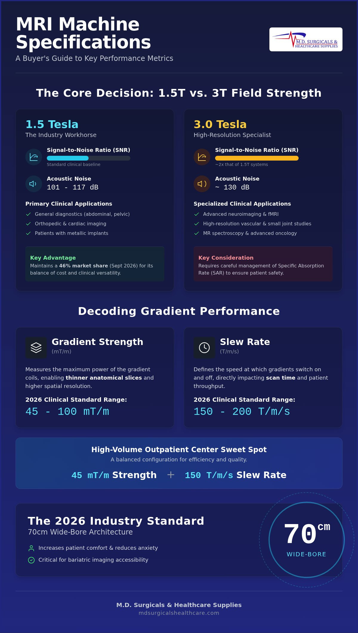
The 1.5T system remains the industry workhorse for general clinical use and orthopedic imaging. It's particularly effective for patients with metallic implants or orthopedic hardware, as it generates fewer susceptibility artifacts than higher field alternatives. Market data from September 2026 indicates that 1.5T systems maintain a 46% market share, proving their continued relevance in diverse clinical settings. These systems are often easier to site and require less extensive radiofrequency shielding than their more powerful counterparts.
In contrast, 3T systems provide approximately double the Signal-to-Noise Ratio (SNR) of 1.5T units. This increased power allows for either significantly faster scan times or much higher spatial resolution, which is essential for capturing minute anatomical details. It's the preferred choice for advanced vascular studies and neuroimaging where precision is paramount. For community clinics in 2026, the 1.5T closed-bore system remains the gold standard due to its balance of clinical versatility and manageable operational costs.
Signal-to-Noise Ratio (SNR) and Image Quality
SNR is the foundational metric for diagnostic confidence; it's the ratio of the desired signal to the background electronic noise. Higher field strength correlates with finer spatial resolution, allowing radiologists to see smaller structures with greater clarity. However, 3T systems require careful management of the Specific Absorption Rate (SAR), which measures the heat energy absorbed by the body. They also tend to be louder, reaching approximately 130 dB compared to the 101 to 117 dB range typical of 1.5T models.
Clinical Applications by Field Strength
Your facility's primary specialty should dictate your choice in mri machine specifications. 1.5T systems are highly effective for routine abdominal, pelvic, and cardiac imaging where motion and susceptibility artifacts are common concerns. Conversely, 3T field strength is a necessity for specialized applications including:
- Functional MRI (fMRI) and brain mapping protocols.
- MR spectroscopy for detailed metabolic analysis.
- High-resolution imaging of small joints or the prostate.
- Advanced neuro-oncology assessments.
Decoding Gradient Performance and Slew Rates
Gradients are the internal magnetic coils responsible for spatial encoding. They create the variation in magnetic field strength that allows the system to identify exactly where signals originate in the body. While the main magnet provides the baseline field, the gradient system provides the resolution. When evaluating mri machine specifications, you must distinguish between gradient strength and slew rate to understand how the system will perform under heavy clinical loads.
Gradient Strength vs. Slew Rate
Gradient strength, measured in millitesla per meter (mT/m), represents the maximum power the gradient can reach. Higher strength allows for thinner anatomical slices and increased spatial resolution. Slew rate, measured in Tesla per meter per second (T/m/s), defines the speed at which the gradients switch on and off. You'll find that interpreting gradient specifications correctly reveals that speed is often more critical than raw power for advanced imaging.
In September 2026, most clinical systems feature gradient strengths between 45 and 100 mT/m and slew rates ranging from 150 to 200 T/m/s. High slew rates are non-negotiable for cardiac imaging, where the system must capture data between heartbeats, and for pediatric cases where scan time must be minimized. For a high-volume outpatient center, the sweet spot for gradient specs typically involves a slew rate of 150 T/m/s paired with a gradient strength of 45 mT/m.
Impact on Patient Throughput
The efficiency of your gradient system directly dictates your daily patient volume. High-duty cycle gradients allow for rapid, repetitive pulsing without the system overheating or requiring "cool-down" periods between sequences. This performance level is essential for advanced sequences like Diffusion Tensor Imaging (DTI), which requires complex spatial encoding. By utilizing high-performance gradients, facilities can often shave five minutes off standard protocols like a routine lumbar spine or knee scan.
If your facility averages 15 scans per day, a five-minute reduction per patient adds 75 minutes of available time to your daily schedule. This allows for additional appointments and significantly boosts your annual revenue. While high-performance gradients increase the initial purchase price, the long-term ROI through increased throughput is substantial. If you're comparing spec sheets from different manufacturers, you can consult our technical specialists to ensure your chosen system meets your specific volume requirements.
Bore Diameter and Coil Technology: Patient Experience Specs
Patient comfort is no longer a secondary consideration; it's an operational necessity that dictates scan success rates. Standard 60 cm bores are increasingly rare in new procurement cycles as healthcare facilities prioritize accessibility. In September 2026, 70 cm wide-bore systems have become the industry standard for high-volume imaging centers. This shift in mri machine specifications directly addresses the two biggest hurdles to patient throughput: claustrophobia and bariatric imaging requirements. A wider opening reduces the frequency of aborted scans and allows for easier positioning of larger patients without sacrificing the field homogeneity required for clear images.
Closed vs. Wide Bore Systems
While some facilities consider "Open MRI" systems using permanent magnets, these typically operate at lower field strengths between 0.3T and 1.0T. These systems often involve significant trade-offs in image quality and require much longer scan times to achieve diagnostic clarity. Wide Bore 1.5T systems offer a superior alternative, providing the comfort of a larger opening with the diagnostic power of a high-field superconducting magnet. To further enhance patient safety and comfort during long sequences, facilities should browse patient monitors that are specifically rated for the MRI environment. These tools allow clinical staff to maintain constant vigilance over physiological signs while the patient is inside the bore.
RF Coil Architecture
Signal reception is just as critical as signal generation. Modern RF (Radio Frequency) coil architecture uses phased-array designs and digital signal transmission to capture data with minimal loss. When reviewing mri machine specifications, you must evaluate the number of channels available in the coil. A 32-channel head coil offers significantly higher signal density than a 16-channel version, which improves parallel imaging performance. This technology allows the system to reconstruct images faster, effectively reducing the time a patient spends in the machine. Lightweight, flexible coils are now preferred because they conform better to patient anatomy, improving both signal reception and patient positioning ease.
The relationship between bore size and coil density is the primary driver of patient satisfaction in 2026. High-channel coils allow for accelerated imaging protocols that, when paired with a 70 cm bore, significantly reduce patient anxiety. This combination ensures that even the most difficult-to-scan populations can be managed with clinical precision. Meticulous evaluation of these hardware specs ensures your facility remains competitive and capable of handling diverse patient demographics.
How to Analyze an MRI Spec Sheet: A Step-by-Step Evaluation
Analyzing mri machine specifications requires a methodical workflow to move beyond marketing rhetoric and into clinical reality. Start by identifying the magnet type. Most modern high-field units are superconducting, but you must verify the helium consumption architecture. Systems like the MAGNETOM Flow platform now utilize as little as 0.7 liters of liquid helium; this significantly reduces the logistical burden of traditional "boil-off" refills. Next, cross-reference the gradient slew rate with your clinical volume. If your facility focuses on cardiac studies, a slew rate approaching 200 T/m/s is necessary to maintain temporal resolution.
Software and AI Integration
By September 2026, AI-driven software is no longer optional for high-ROI facilities. Deep Learning Reconstruction (DLR) algorithms can reduce image noise and shorten scan times by up to 50%. These packages often represent a separate capital expenditure between $50,000 and $200,000. Ensure the system maintains full DICOM compatibility for seamless integration with your existing Picture Archiving and Communication System (PACS). For facilities performing advanced cardiac assessments, you should also explore ECG machines that offer compatible gating triggers for synchronized imaging.
Logistical and Environmental Specs
Physical site compatibility often presents the most significant hurdle during installation. You must audit the fringe field requirements, specifically the 5-Gauss line, to ensure proper room shielding for safety. Floor loading is another critical metric; some 3.0T systems exceed 7,000 kg, which may require structural reinforcement for second-story placements. Verify the power requirements and cooling capacity of your facility's HVAC system to prevent unplanned operational downtime. Finally, confirm the hardware’s FDA 510(k) clearance and accreditation status to ensure compliance with clinical standards. A meticulous audit of these mri machine specifications prevents costly site modifications after the equipment arrives.
Sourcing Verified MRI Equipment for Global Healthcare
Procuring high-value diagnostic hardware requires a level of due diligence that extends beyond reviewing mri machine specifications. Working with a licensed distributor is the only way to ensure that your equipment meets stringent international standards and manufacturer performance benchmarks. M.D. Surgical & Healthcare Supplies acts as a strategic partner by verifying the provenance and accreditation of every system within our global network. This meticulous verification process protects your institution from the operational risks associated with grey-market hardware or uncertified imaging systems.
The Role of the Licensed Distributor
Navigating the regulatory landscape of different international jurisdictions is a complex task that demands professional expertise. A licensed partner ensures that every component complies with the standards set by the Food and Drug Administration (FDA) and other relevant health authorities. Choosing a single-source provider allows you to streamline your procurement by sourcing both advanced imaging systems and critical PPE consumables, such as N95 respirators (NIOSH-approved). Maintaining supply continuity in diagnostic imaging is the most effective way to prevent clinical bottlenecks and ensure that your facility remains a dependable pillar of your local healthcare ecosystem.
Global shipping for an MRI scanner involves sophisticated logistical planning, including the use of climate-controlled containers and continuous vibration monitoring. Our logistical framework is designed to manage these complexities, ensuring that your machinery arrives in peak condition within the verified lead times for September 2026. Institutional buyers and large hospital networks can leverage wholesale pricing structures that offer a clear financial advantage over traditional retail procurement. These cost efficiencies allow for a more sustainable Return on Investment (ROI) while maintaining the highest levels of clinical precision.
Next Steps in Your Procurement Journey
Preparing your facility for the arrival of new diagnostic equipment requires early coordination with structural engineers and specialists in Heating, Ventilation, and Air Conditioning (HVAC). You must ensure that your site meets the specific mri machine specifications regarding floor loading and electromagnetic shielding before the delivery date. Requesting a tailored quote is the first step toward securing a system that aligns with your facility's patient volume and clinical specialization. To begin this process, Contact our supply specialists for a comprehensive consultation. We provide the administrative clarity and technical support necessary to navigate the global medical supply chain with absolute confidence.
Securing Your Facility's Imaging Infrastructure
Selecting the right diagnostic system requires a precise alignment between clinical goals and technical hardware. You've now mastered the essential mri machine specifications needed to evaluate 1.5T and 3T systems while understanding how gradient slew rates dictate your facility's daily patient volume. By prioritizing 70 cm wide-bore architecture and AI-driven reconstruction, your institution can ensure both diagnostic accuracy and patient comfort. These technical parameters aren't just engineering metrics; they're the primary drivers of your operational ROI.
Working with a licensed distributor provides the logistical stability and equipment accreditation necessary for a successful global deployment. M.D. Surgical & Healthcare Supplies specializes in sourcing verified medical hardware and offering competitive institutional wholesale pricing to hospital networks. We manage the complexities of international shipping and regulatory compliance so your clinical staff can focus on patient care. Our expertise ensures that your high-value machinery arrives verified, accredited, and ready for installation.
Our team is ready to help you prepare your facility for the next generation of medical imaging. We look forward to supporting your procurement journey with professional authority and logistical precision.
Frequently Asked Questions
What is the difference between 1.5T and 3T MRI machine specifications?
3T systems offer approximately double the signal to noise ratio of 1.5T units, enabling higher resolution or faster scans. While 3T is superior for neuroimaging and spectroscopy, 1.5T remains the industry standard for general clinical use and imaging near metal implants. Choosing between these mri machine specifications depends on your facility's specialty and budget. High end imaging systems generally fall within the $50,000–$500,000+ price range depending on field strength and configuration.
How does the MRI bore size affect image quality and patient comfort?
Bore size directly impacts the patient experience and the homogeneity of the magnetic field. A 70 cm wide bore system is the 2026 market standard for reducing claustrophobia and accommodating bariatric patients. While larger bores can make maintaining a perfectly uniform magnetic field more challenging, modern shim technology ensures that image quality remains high. Selecting a wider bore often leads to fewer aborted scans and improved patient throughput in high volume imaging centers.
What is a slew rate in an MRI scanner and why does it matter?
Slew rate measures the speed at which the gradient magnetic fields can be switched on and off. It's calculated in Tesla per meter per second (T/m/s). Higher slew rates allow for faster imaging sequences and thinner anatomical slices, which are critical for cardiac and pediatric protocols. In the United States, the FDA limits slew rates to 200 T/m/s to prevent peripheral nerve stimulation. This metric is a vital part of mri machine specifications for high performance facilities.
Can a 1.5T MRI machine perform advanced neurological scans?
Yes, a 1.5T system can perform most advanced neurological scans, though it may require longer acquisition times than a 3T unit. It remains highly effective for routine brain imaging, stroke evaluation, and spinal assessments. For specialized research, functional MRI, or high resolution spectroscopy, a 3T magnet is often preferred for its superior signal density. Most community clinics find that a 1.5T system provides the necessary versatility for a broad range of neurological clinical cases.
What are the power and cooling requirements for a modern MRI scan machine?
Modern systems require a stable three phase power supply and a dedicated HVAC system to manage the heat generated by gradient coils. Superconducting magnets typically use liquid helium for cooling, though 2026 technology favors zero boil off systems that recycle helium internally. You must also account for the 5 Gauss exclusion zone to ensure the safety of staff and patients with metallic implants. Proper site preparation prevents operational downtime and ensures the long term reliability of your imaging hardware.
How do I verify the accreditation of used MRI equipment for sale?
You should only source equipment through a licensed distributor that provides a clear record of provenance and accreditation. All products at M.D. Surgical & Healthcare Supplies are accredited and verified before sale to ensure they meet international safety standards. Request documentation of FDA 510(k) clearance and a comprehensive service history. Working with a partner that has a global network across four regional hubs ensures that the hardware complies with local regulatory requirements.
What is the lifespan of a superconducting MRI magnet in 2026?
A well-maintained superconducting magnet can last for 20 years or more if the vacuum integrity and cooling systems are preserved. While the software and gradient electronics may require upgrades every five to eight years, the main magnet remains a long term capital asset. Regular maintenance and the use of helium saving technologies extend the operational life of the system. Institutional buyers should factor this longevity into their long term ROI calculations when comparing high end imaging hardware.
Are there portable MRI machine specifications suitable for clinics?
Portable or low field MRI systems, such as 0.55T High V units, are increasingly available for point of care use. These systems have a smaller physical footprint and reduced shielding requirements, making them ideal for emergency departments or smaller clinics. While they don't match the signal intensity of 1.5T or 3T magnets, they excel at lung imaging and scanning patients with metal implants. These systems represent a growing segment of the diagnostic market for facilities with space constraints.

